Pelvic Floor Muscles Ct Scan
The Pelvis Radiology Key
Http Pdf Posterng Netkey At Download Index Php Congress Ecr2014 Module Get Pdf By Id Poster Id 119484

Above Shows A Number Of Possible Measurements Using Mri Imaging A Download Scientific Diagram
Ct Abdomen Pelvis Lower Axial Labeling Questions Radiology Case Radiopaedia Org

Figure 3 From Obturator Hernia Semantic Scholar
Presentation1 Pptx Ct Normal Anatomy Of The Abdomen And Pelvis
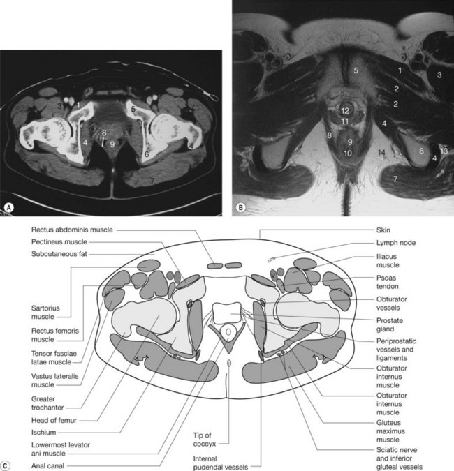
2 psoas muscle 5 femur 6 obturator internus muscle 8 pubis 9 ischium 11 levator ani muscle 29 anus 33 urethra 35 penis 50.
Pelvic floor muscles ct scan. When are they useful. An abdominal ct takes pictures of your abdomen. If your doctor recommends a pelvis ct scan you likely have questions. Radiologic study that helps rule out other medical conditions that may have similar symptoms to prolapse.
Abdominal ct scans also called cat scans are a type of specialized x ray. Male abdomen and pelvis ct scan form no 19. 2 psoas muscle 4 sacrum 6 obturator internus muscle 13 ureter 14 bladder 22 small bowel 27 sigmoid colon 28 rectum 30 vas deferens 31 seminal vesicles. If you notice prolapsed pelvic organs.
A pelvic ct scan takes pictures of your pelvis the area between your hips. A ct scan uses x rays to look at bones muscles body organs and blood vessels. Anatomy of the abdomen and male pelvis using cross sectional imaging ct interactive atlas of human anatomy we created an anatomical atlas of abdominal and pelvic ct which is an interactive tool for studying the conventional anatomy of the normal structures based on a multidetector computed tomography. Radiologic study that assesses the muscles organs and support of the pelvic floor and helps to evaluate how the pelvic floor functions with straining.
What can these scans do. A pelvic mri scan uses magnets and radio waves to help your doctor see the bones organs blood vessels and other tissues in your pelvic region the area between your hips that holds your. Normally these muscles and the tissues surrounding them keep the pelvic organs in place. This enables images to be taken of the bladder urethra water pipe vagina cervix uterus womb perineum anal canal and pelvic floor muscles.
Ct scan of abdomen and pelvis. They help your doctor see the organs blood vessels and bones in your abdomen. This scan can help determine the cause of urinary incontinence. We ll explain why your doctor may.
The pelvic floor is a group of muscles that form a kind of hammock across your pelvic opening. A pelvic scan will be carried out before we do the pelvic floor scan. Who they can help.

Predictive Role Of Measurement Of Pelvic Floor Muscle Thickness With Static Mri In Stress And Mixed Urinary Incontinence Semantic Scholar

State Of The Art An Integrated Approach To Pelvic Floor Ultrasonography Santoro 2011 Ultrasound In Obstetrics Amp Gynecology Wiley Online Library
Computed Tomography Evaluation Of The Piriformis Muscle Axial Computed Download Scientific Diagram

Pelvis And Abdomen Radiology Key

Pelvic Floor Disorders Pelvic Girdle Pain And Symphysis Pubis Dysfunction Following Sports Injury Caring Medical

Pelvic Muscle Attachments A Classic Exam Question 1 Multiple Abdominal Muscles Iliac Crest Medical Anatomy Anatomy Medical

Magnetic Resonance Imaging Of The Female Pelvic Floor Radiology Key

Epos Trade

Pelvis Radiographic Anatomy Wikiradiography Radiology Student Medical Knowledge Radiography
Which Cancers Can A Pelvic Ct Scan Detect American Health Imaging
Https Www Abdominalradiology Org Resource Resmgr Education Dfp Pelvicfloor Anatomy Pelvic Floor Imaging And Ana Pdf

