Pelvic Floor Mri Radiographics

Mri Pelvis Anatomy Free Male Pelvis Axial Anatomy

Magnetic Resonance Imaging Of The Pelvic Floor Sciencedirect
Https Www Abdominalradiology Org Resource Resmgr Education Dfp Pelvicfloor Mri Imaging Mri Of Pelvic Floor Dysfunct Pdf
Https Www Ajronline Org Doi Pdf 10 2214 Ajr 07 7096
Http Pdf Posterng Netkey At Download Index Php Module Get Pdf By Id Poster Id 117382

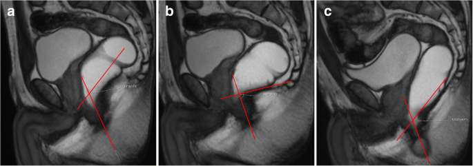
Image Based Measurements For Evaluation Of Pelvic Organ Prolapse
Mr imaging based assessment of the female pelvic floor.
Pelvic floor mri radiographics. Magnetic resonance imaging of pelvic floor relaxation. Bertschinger km hetzer fh roos je treiber k marincek b hilfiker pr. Mri of pelvic floor dysfunction. Imaging can play an additional role in the postoperative setting in the evaluation.
Paraurethral ligaments arrowheads in a which arise from the lateral wall of the urethra u. Role of static and dynamic mr imaging in surgical pelvic floor dysfunction. The authors review the pelvic floor anatomy describe the mr imaging protocol used in their institutions survey common mr imaging findings in the presence of pelvic floor weakness and highlight key details that radiologists should provide surgeons to ensure effective treatment and improved outcomes. Ajr am j roentgenol.
The periurethral ligaments arrows which arise from the pu borectalis muscle. Mri of pelvic floor dysfunction esur and esgar recommendations. Mr imaging of the female pelvic floor in the supine and upright positions. The anatomy and biomechanics of genital prolapse.
Dynamic mr imaging of the pelvic floor performed with patient sitting in an open magnet unit versus with patient supine in a. Magnetic resonance imaging of pelvic floor disorders. Pelvic floor failure is a common disorder that affects 23 7 of women in the united states with a prevalence of 9 7 49 7 that increases with age one in nine women will undergo an invasive procedure for treatment of urinary incontinence or pelvic organ prolapse with 30 requiring additional surgery for symptom recurrence by 80 years of age. Boyadzhyan l raman ss raz s.
Top magn reson imaging. Imaging in the perioperative setting can be used as an objective measure after pelvic floor intervention to document anatomic and functional changes 8 12. Clin obstet gynecol 1993. Colaiacomo mc masselli g polettini e et al.
Normal female pelvic floor anatomy. 36 stoker j halligan s bartram c. Axial t2 weighted mr images show the ligaments that support the female urethra at superior a and inferior b levels. Mr imaging of pelvic floor.
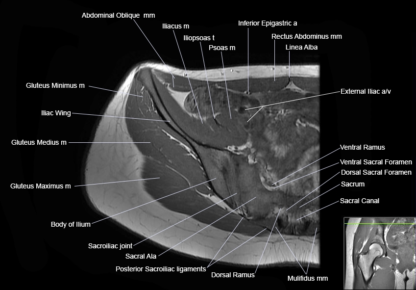
Practical mr imaging of female pelvic floor weakness. Role of static and dynamic mr imaging in surgical pelvic floor dysfunction. The female pelvic floor is composed of the vulva levator ani muscle deep to it and the hollow viscera urethra vagina and rectum that penetrate through the levator ani at the midline 7 8 the supporting framework is the pelvic bony ring pubic rami ischium ilium sacrum and coccyx. 28 fielding jr versi e mulkern rv lerner mh griffiths da jolesz fa.
However imaging findings may not always correlate well with the clinical findings and symptoms 13 14.
Dynamic Magnetic Resonance Imaging Of The Female Pelvic Floor A Pictorial Review Springerlink
Mri Hip Anatomy

Mr Defecating Proctography Radiology Reference Article Radiopaedia Org

Dynamic Magnetic Resonance Imaging For Assessment Of Minimally Invasive Pelvic Floor Reconstruction With Polypropylene Implant European Journal Of Radiology
Mr Based Imaging Of Female Pelvic Floor

Dynamic Mr Imaging Of The Pelvic Floor A Pictorial Review Radiographics

Mri Anatomy Of Hip Joint Free Mri Axial Hip Anatomy Hip Anatomy Mri Pelvis Anatomy

Constipation Mri Wikidoc

Pelvic Floor Dysfunction Failure Mri Radiology Albums Facebook
Pdf Magnetic Resonance Imaging Of Pelvic Floor Dysfunction Joint Recommendations Of The Esur And Esgar Pelvic Floor Working Group

Translabial Us And Dynamic Mr Imaging Of The Pelvic Floor Normal Anatomy And Dysfunction Radiographics

Mri Female Pelvis Anatomy Axial Image 16 Pelvis Anatomy Pelvis Rectus Abdominis Muscle
Global Pelvic Floor Descent During Evacuation Radiology Case Radiopaedia Org