Pelvic Floor Emg Testing

Reliability Of Pelvic Floor Muscle Electromyography Tested On Healthy Women And Women With Pelvic Floor Muscle Dysfunction Sciencedirect
Low Back Pain And The Pelvic Floor Musculoskeletal Key

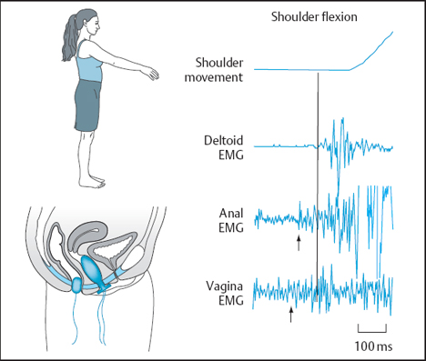
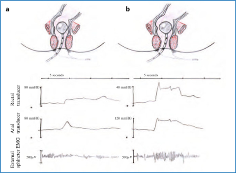
Contraction Of The Pelvic Floor Muscles During Abdominal Maneuvers Archives Of Physical Medicine And Rehabilitation

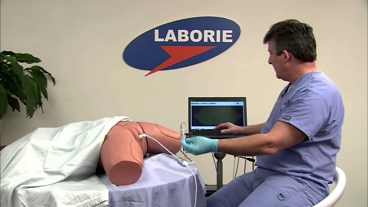
Srs Medical Aware Wireless Emg Incontinence Diagnostics Treatments
Biofeedback And Pelvic Floor Disorders Abdominal Key

Surface Electromyography Evaluation Etiology

Electrodiagnostic testing emg of the pelvic floor this testing evaluates nerve function of the pelvic floor.
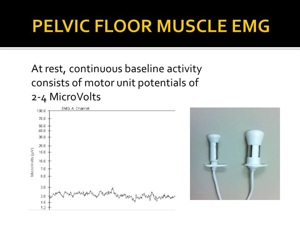
Pelvic floor emg testing. A uroflow test can show how well you can empty your bladder. The unit of measure is in microvolts µv. Pelvic floor muscle emg is most commonly recorded as part of urodynamic studies to obtain information about the kinesiological behavior of pelvic floor structures during. Emg determines the pelvic floor s muscle response to a series of small electrical impulses.
Emg studies used to evaluate pelvic floor disorders can be broadly separated into two categories kinesiological emg kemg and motor unit emg. Pfm emg has shown good reliability in healthy women. If your flow of urine is weak or if you have to stop and start as you urinate it can point to pelvic floor dysfunction. Heitner 69 concluded that surface emg was superior to vaginal palpation in assessment of all variables other than lift.
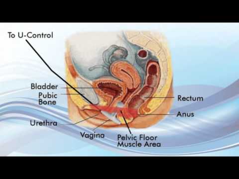
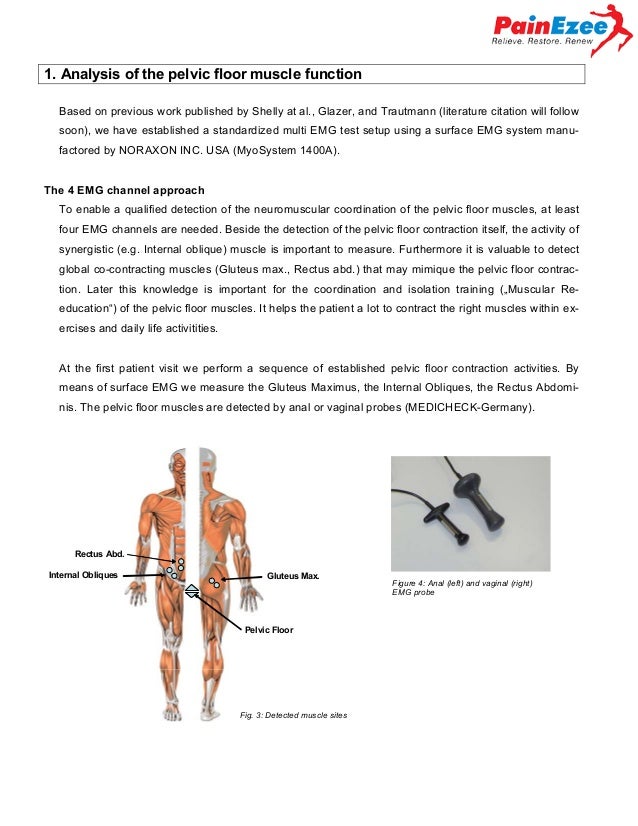
This test is not painful. Pelvic floor muscle location. Pelvic floor muscle surface emg or biofeedback manual muscle testing internal or external postural exercises internal and external massage myofascial release relaxation techniques and diaphragmatic breathing development of an individualized home exercise. 31 kemg is used simply to assess the activity or inactivity of a muscle usually the urethral or anal sphincter.
However its reliability has not been tested in women with pfm dysfunction. Electromyography emg is a well established method to quantify the relative pelvic floor muscle pfm activity. Of the many emg techniques only a few have use for the study of pelvic floor disorders. Electromyography emg the term electromyography is a general term that refers to methods of studying electrical activity of muscle.
Emg is a test to determine if the nerves that supply your sphincter muscle are intact and that all your muscles relax and contract as they should. It is used in conjunction with physiologic tests such as urodynamics and anal manometry. Depending on your condition additional tests such as anal manometry defecography anal ultrasound and pelvic magnetic resonance imagery. These tests are performed by measuring the electrical activity with a small sponge in the anus or by stimulating the nerve with a small electrode.
Your provider may order this test if you have problems while urinating.
Urostym Pelvic Floor Rehabilitation System Youtube
Pelvic Floor Biofeedback For Incontinence Youtube

Flow Electromyography Emg And Pressure Profiles Laborie A Flow Emg Download Scientific Diagram

Pdf Pelvic Muscle Rehabilitation A Standardized Protocol For Pelvic Floor Dysfunction
Women S Health And Education Center Whec Urodynamic Assessment Electromyography
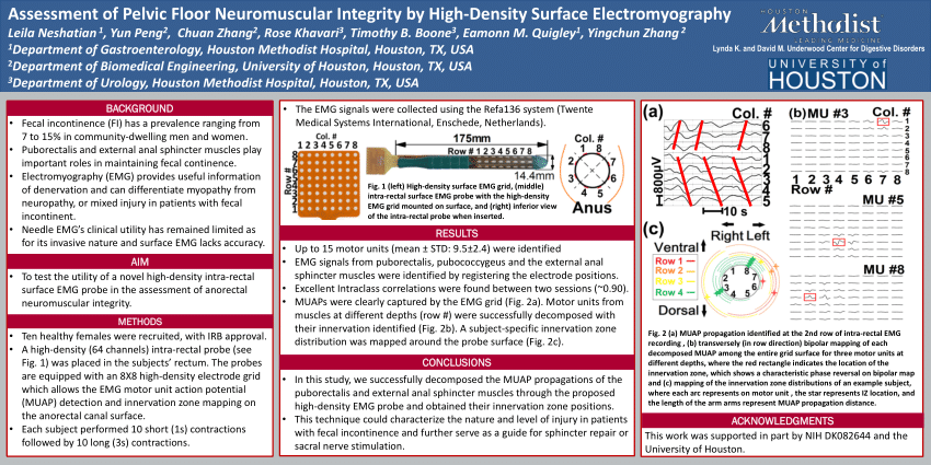
Pdf Assessment Of Pelvic Floor Neuromuscular Integrity By High Density Surface Electromyography
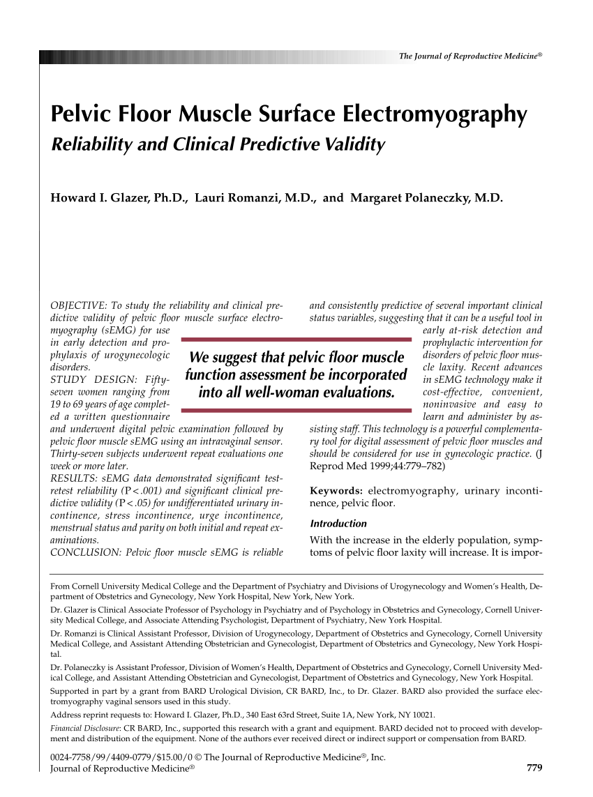
Pdf Pelvic Floor Muscle Surface Electromyography Reliability And Clinical Predictive Validity
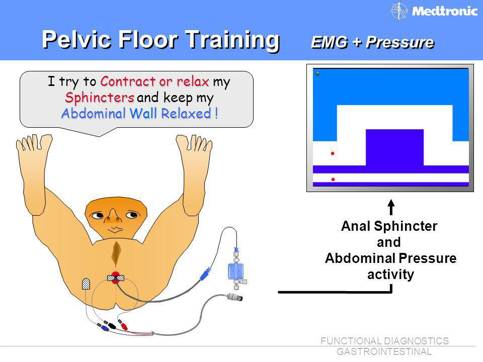
Functional Diagnostics Gastrointestinal What Is Biofeedback Q Provides The Patient Immediate Auditory And Or Visual Information About Physiological Process Ppt Download

Apparatus With Multiple Functions Measurement Of Pelvic Floor Muscle Download Scientific Diagram
Pelvic Health In Women Beth Lonberger Aprn Family Nurse Practitioner Ppt Video Online Download

A New Device For Simultaneous Measurement Of Pelvic Floor Muscle Activity And Vaginal Blood Flow A Test In A Nonclinical Sample The Journal Of Sexual Medicine
Zmpczm019000 11 03 Emg Based Evaluation Therapy Concept For Pelvic

Pelvic Floor Muscles Salli

